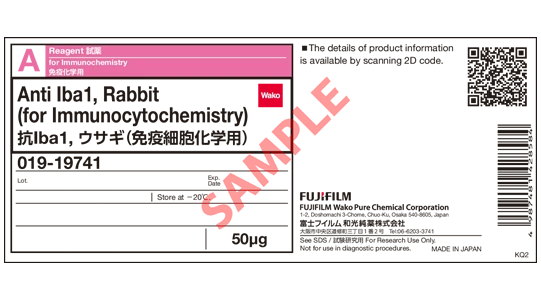
Anti Iba1, Rabbit (for Immunocytochemistry)
| Grade : | for Immunochemistry |
| Storage Condition : | Keep at -20 degrees C. |
| Source : | Rat |
| Cross‒Reactivity : | Mouse, Rat |
| Host : | Rabbit |
| Application : | ICC, IHC(Frozen) |
| Catalogue/Product Number | Package Size | COA Download |
|---|---|---|
|
019-19741
|
50ug |

Document
Overview
Iba1 (Ionized calcium-binding adapter molecule1) is an approximately 17 kDa calcium-binding protein. It is used as a microglial marker because it is expressed specifically in microglia in the central nervous system1). It is expressed in both resting and activated microglia, but is reportedly expressed more highly in activated microglia2). It is also expressed in macrophages in peripheral tissues and is known as AIF-1 (Allograft inflammatory factor-1). Iba1 binds to F-actin in cells to form actin bundles. The formation of actin bundles is thought to be required for the membrane ruffling observed during cell migration and phagocytosis3).
Fujifilm Wako’s "Anti Iba1, Rabbit (for immunocytochemistry)" (Product Number 019-19741), which allows even microglia processes to be stained by immunohistochemical staining, is used by researchers all over the world as a microglia marker antibody standard.
Features
- Enables even the processes of microglia to be immunohistochemical-stained*.
- Relatively small amount needed, with concentrations ranging from 1:500 to 1:1,000.
* The result of staining depends on sample condition. Staining performance is not guaranteed.
Antibody Information
| Clonality | Polyclonal |
|---|---|
| Antigen | Synthetic peptide (Iba1 C-terminal sequence) |
| Host | Rabbit |
| Reactivity* | Mouse, Rat |
| Concentration (mg/mL) | 0.5 - 0.7 |
| Applications | Immunohistochemistry (Frozen Section) 1:500 - 1,000 Immunocytochemistry 1:500 - 1,000 |
* Experience with humans4) dogs5), cats6), pigs7), marmosets8), zebrafish9), etc. has been reported.
Standard Protocol for Immunohistochemistry of Microglia using Iba1 antibody
"Anti Iba1, Rabbit (for Immunocytochemistry)" is an excellent microglial marker antibody since it can stain even microglial processes. Here, we describes the protocol and points to note when performing microglial immunohistochemistry. Frozen sections of mouse brain and a fluorescent dye are used as an example.
1.Preparation of tissue sections
1-1. Mice are perfused and fixed in 4% paraformaldehyde-phosphate buffer.
[Point] Samples without perfusion fixation or with inadequate fixation result in poor staining. Perfusion fixation with 4% paraformaldehyde-phosphate buffer is recommended.
1-2. Replace with sucrose, and prepare frozen blocks.
1-3. Prepare 50 µm-sections in thickness using a microtome.
[Point] Recommended thickness of tissue sections is 20-50 µm.
2.Washing - Blocking
2-1. Wash with 0.3% TritonX-100/PBS 3 times for 5 minutes each.
2-2. Block in 1% BSA, 0.3% TritonX-100/PBS for 2 hours at room temperature.
[Point] If background is high, try extending the incubation time for blocking or changing the blocking solutions.
The following blocking solutions can also be used:
· 1% BSA, 0.3% Tween-20/PBS
· 3% normal serum of the host of the secondary antibody
3.Primary antibody reaction
3-1. Add Anti Iba1, Rabbit (for immunohistochemistry) to 1% BSA, 0.3% TritonX-100/PBS at 1:1,000 dilution.
[Point] If the staining is weak, increase the antibody concentration. If the background is high, decrease the antibody concentration. Recommended dilution is 1:500-1,000.
3-2. Incubate overnight at 4°C.
[Point] Although it depends on the sample, rat cerebellum has been successfully stained with just 2 hours of incubation.
4.Washing
4-1. Wash with 0.3% TritonX-100/PBS 3 times for 5 minutes each.
5.Secondary antibody reaction
5-1. Add fluorescent-labeled anti-rabbit IgG antibody (e.g. Jackson ImmunoResearch, Product No. 111-545-144), to 1% BSA, 0.3% TritonX-100/PBS at 1:1,000 dilution.
[Point] If the staining is weak, increase the antibody concentration. If the background is high, decrease the antibody concentration. Recommended dilution is 1:500-1,000.
5-2. Incubate for 2 hours at room temperature.
[Point] If the background is high, shorten the incubation time for the secondary antibody. Recommended incubation time is 1-2 hours.
6.Washing
6-1. Wash with 0.3% TritonX-100/PBS 3 times for 5 minutes each.
[Point] If the background is high, increase the number of washes.
7.Mounting
7-1. Mount the sections in mounting media.
8.Observation
8-1. Observe the sections under a fluorescence microscope or a confocal microscope.
If microglia do not stain well using the techniques described at left, perform antigen retrieval after sectioning using one of the following:
(A) Citrate buffer (pH 6.0) for 9 minutes at 90°C
(B) TE bufferi (pH 9.0) for 9 minutes at 90°C
Other troubleshooting is also available in the FAQ. .
Application Data
Immunohistochemistry (Mouse)
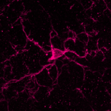
| Species | Mouse |
| Site | Cerebellum |
| Sample | Frozen section |
| Antibody concentration | 1:1,000 |
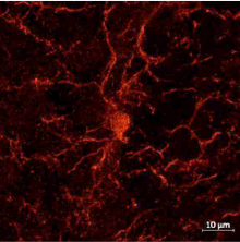
| Species | Mouse |
| Site | Nucleus accumbens core |
| Sample | Slice section |
| Antibody concentration | 1:500 |
| Data by courtesy of | Dr. Hagiwara, Mr. Yamazaki Tokyo University of Science |
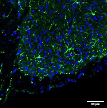
| Species | Mouse |
| Site | Spinal cord |
| Sample | Frozen section |
| Antibody concentration | 1:500 |
| Data by courtesy of | Dr. Hagiwara, Mr. Sawada Tokyo University of Science |
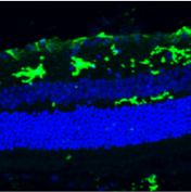
| Species | Mouse |
| Site | Retinal whole mount |
| Antibody concentration | 1:1,000 |
| Data by courtesy of | Prof. Lieve Moons KU Leuven Belgium |
Immunohistochemistry (Rat)
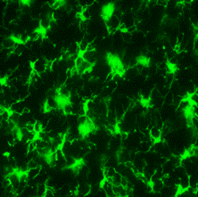
| Species | Rat |
| Site | Cerebral cortex |
| Sample | Frozen section |
| Antibody concentration | 1:1,000 |
| Data by courtesy of | Dr. Sanagi, Dr. Manabe, Dr. Ichinohe, Dr. Kohsaka National Center of Neurology and Psychiatry |
Overview / Applications
| Outline | This product is for research use only. Do not administer it to human. Calcium ions are known to be one of the most important signal mediators in all cells including central nervous system (CNS) cells. Calcium ions exert their signaling activity through association with various calcium binding proteins, many of which are classified into a large protein family, the EF hand protein family.Iba1 is a 17-kDa EF hand protein that is specifically expressed in macrophages/ microglia and is upregulated during the activation of these cell. Wako has launched rabbit polyclonal antibodies were raised against a synthetic peptide corresponding to the Iba1 carboxy-terminal sequence, which was conserved among human, rat and mouse Iba1 protein sequences. Rabbit Anti Iba1 antibody is raised a synthetic peptide corresponding to C-terminus of Iba1. Purified by the antigen affinity chromatography from rabbit antisera and prepared in TBS solution. Contains no preservatives and stabilizers. Reactive with mouse and rat Iba1. Working Concentration: Immunocytochemistry 1-2ug/mL Immunohistotochemistry 0.5-1ug/mL Storage: Store at -20C. After opening, aliquot contents and freeze at -20C. Package Size:50ug (100uL) References:Imai, Y., Ibata, I., Ito, D., Ohsawa, K. and Kohsaka, S.: Biochem. Biophys. Res. Commun., ;224, 855 (1996). Ito, D., Imai, Y., Ohsawa, K, Nakajima, K, Fukuuchi, Y. and Kohsaka, S.: Brain Res. Mol. Brain Res., 57, 1 (1998). Ohsawa, K., Imai, Y., Kanazawa, H., Sasaki, Y. and Kohsaka, S.: J. Cell Sci., 113, 3073 (2000). Sasaki, Y., Ohsawa, K., Kanazawa, H., Kohsaka, S. and Imai, Y.: Biochem. Biophys. Res. Commun., 286, 292 (2001). Kanazawa, H., Ohsawa, K., Sasaki, Y., Kohsaka, S. and Imai, Y.: J. Biol. Chem.,. 277, 20026(2002). |
|---|
Antibody Information
| Antigen | Iba1 |
|---|---|
| Antigen Alias | Iba1 Iba 1 |